The human brain is an incredibly complex organ made up of various parts, each with its own specific functions and responsibilities. Together, these structures control everything from our thoughts and emotions to our basic bodily functions like breathing and heart rate. Some parts are responsible for higher-order processes like memory and decision-making, while others regulate essential functions that keep us alive. Understanding the largest components of the brain and their roles helps us appreciate how this remarkable organ operates as a whole. In the following, we’ll explore the 20 largest parts of the brain, explaining what they do and how they contribute to our everyday life.
Cerebrum
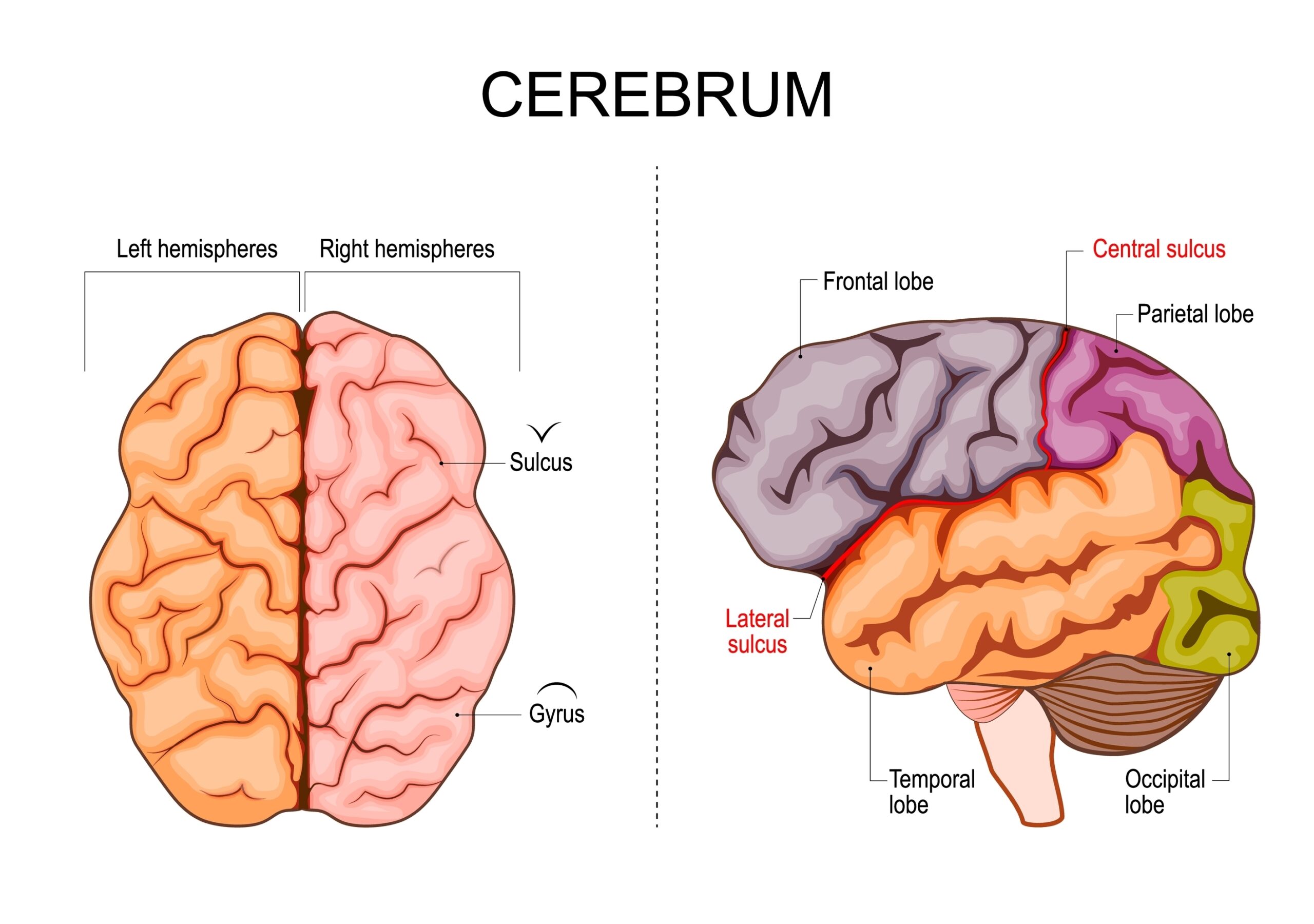
The cerebrum is the largest part of the brain, occupying about 85% of its total mass. It is divided into two hemispheres, each responsible for different cognitive and motor functions. These hemispheres are further divided into four lobes: frontal, parietal, temporal, and occipital. The cerebrum is responsible for higher brain functions, such as thought, reasoning, and emotion. It also controls voluntary muscle movements and processes sensory data. With a surface area of approximately 2,000 square centimeters (due to its many folds called gyri and sulci), the cerebrum is highly complex. Beneath the cortical layers of the cerebrum lie deep structures, including the basal ganglia, which play crucial roles in motor control.
Cerebellum
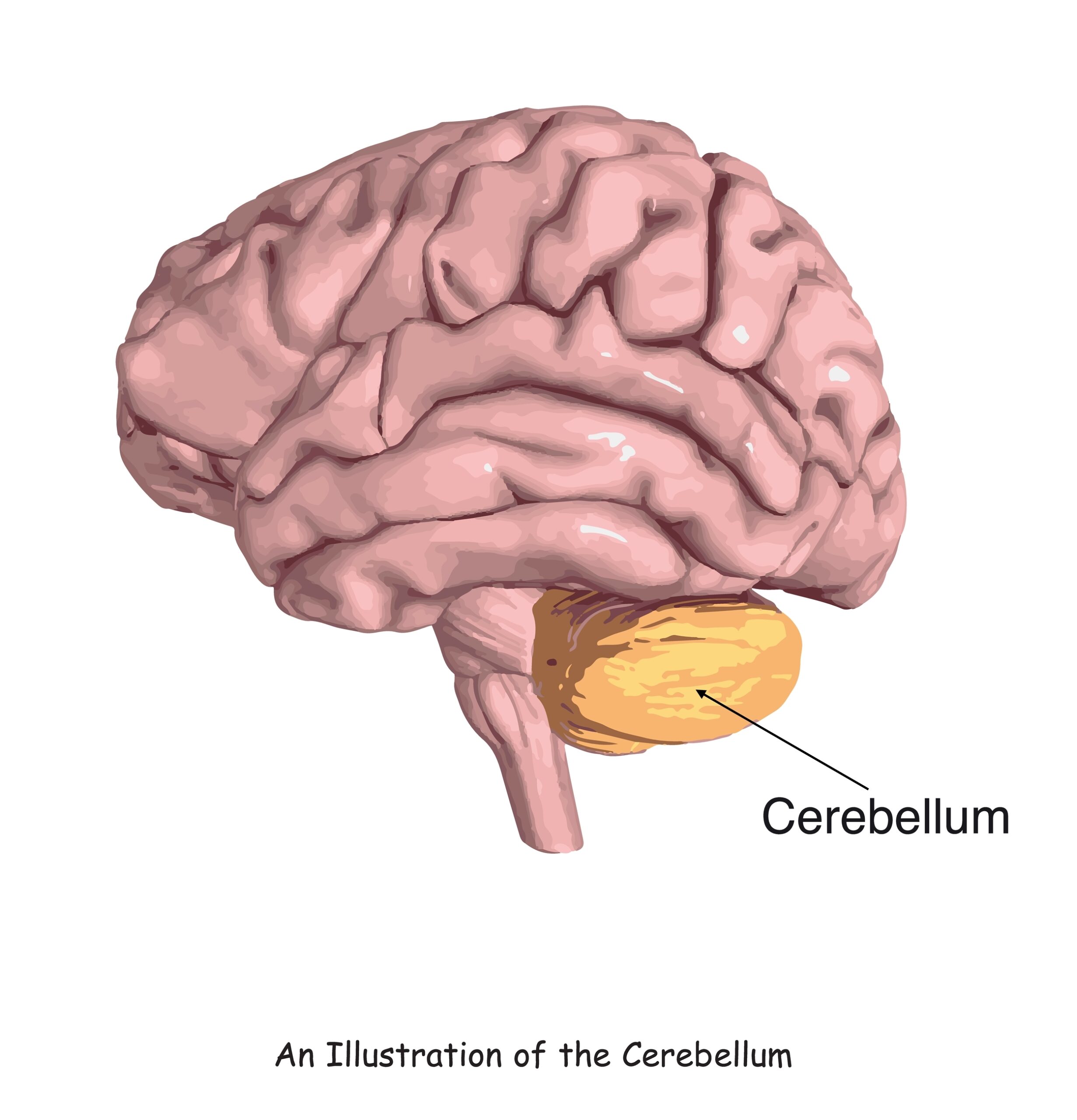
Located beneath the cerebrum at the back of the brain, the cerebellum weighs roughly 150 grams and makes up about 10% of the brain’s volume. Despite its smaller size, it contains more than half the neurons in the entire brain. The cerebellum is primarily responsible for fine-tuning motor movements, balance, and coordination. It works unconsciously, adjusting posture and muscle tone without the need for conscious thought. This structure is also involved in learning motor skills and adapting movements to new situations. Its intricate folds allow it to process vast amounts of information from the body and environment. Damage to the cerebellum can result in difficulties with balance, coordination, and precision in movement.
Frontal Lobe
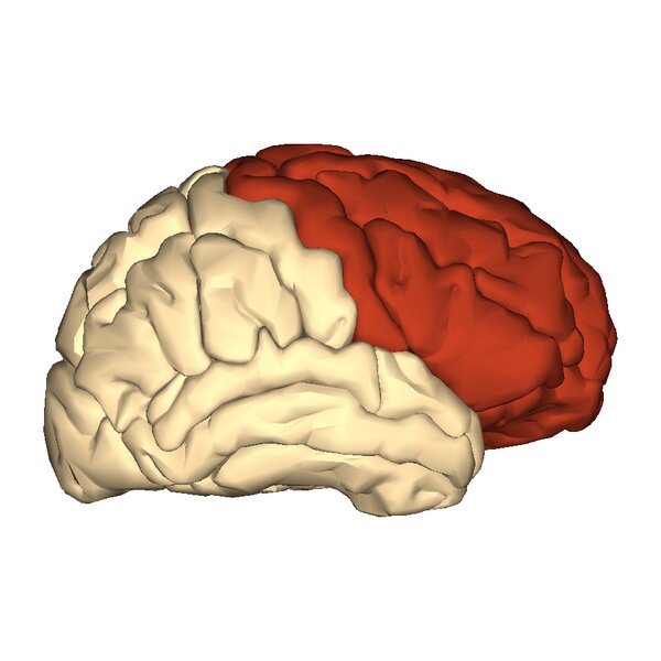
The frontal lobe is the largest lobe of the brain, located in the front of each cerebral hemisphere, and is responsible for many of the brain’s higher cognitive functions. It extends from the forehead to the middle of the head and is involved in decision-making, problem-solving, control of purposeful behaviors, and social interaction. This lobe also houses the primary motor cortex, which controls voluntary movements. Its role in regulating emotions, impulse control, and attention makes it essential for social functioning. The frontal lobe is about one-third of the entire cerebral cortex in humans. Damage here can result in profound changes in personality and the ability to plan or initiate actions.
Parietal Lobe
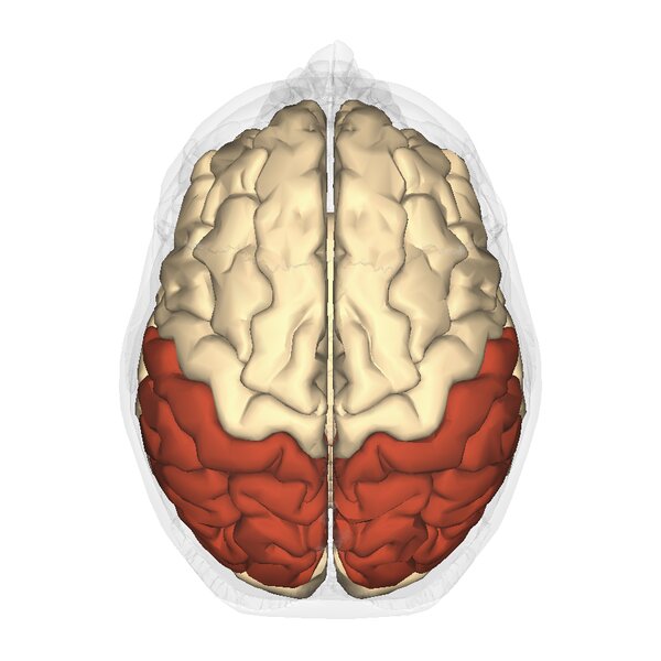
The parietal lobe sits behind the frontal lobe, and its size is substantial, spanning a significant portion of the brain’s surface area. This lobe processes sensory information from the body, particularly touch, temperature, and pain. The primary somatosensory cortex, located here, receives and interprets signals from various parts of the body. It is also involved in spatial orientation and navigation, integrating sensory input to help us understand our physical surroundings. Furthermore, the parietal lobe helps coordinate hand-eye movements and the manipulation of objects. Its overall surface area covers about 600 square centimeters, making it essential for sensory integration. Deficits in this region can lead to difficulties in spatial awareness and body coordination.
Temporal Lobe
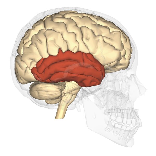
The temporal lobe is positioned beneath the lateral fissure on both sides of the brain and makes up around 22% of the total brain volume. Its functions include processing auditory information, memory formation, and the interpretation of visual stimuli. The hippocampus, an essential structure for memory, is housed within the temporal lobe. In addition to processing sounds, the temporal lobe plays a key role in understanding language and speech. It is also involved in encoding and recalling visual memories. Damage to this area can result in language deficits, memory problems, and difficulties recognizing objects. The temporal lobe covers about 400 square centimeters of cortical surface area.
Occipital Lobe
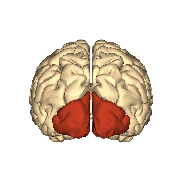
Located at the very back of the brain, the occipital lobe is the primary visual processing center. Despite being the smallest of the four main lobes, it is crucial for interpreting visual stimuli from the eyes. It contains the primary visual cortex, which receives and processes information about color, light, and movement. The occipital lobe is also responsible for visual perception and the recognition of objects. Its surface area is approximately 300 square centimeters. Damage to this region can lead to visual hallucinations, difficulties in recognizing objects, or even blindness. This lobe communicates closely with the temporal and parietal lobes to interpret visual information.
Thalamus
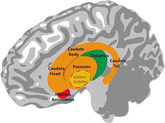
The thalamus is a small, egg-shaped structure located deep within the brain and serves as the brain’s main relay station. It is about 3 centimeters in length and weighs around 0.25% of the brain’s total mass. Sensory information from the body (except smell) passes through the thalamus before being directed to the appropriate areas of the cerebral cortex. The thalamus is involved in regulating consciousness, alertness, and sleep. Its central location allows it to connect various parts of the brain, coordinating motor signals and emotional responses. Damage to the thalamus can result in sensory processing disorders or difficulties in attention and consciousness.
Hypothalamus
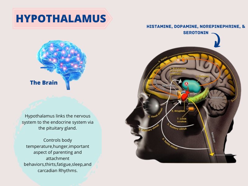
The hypothalamus is a small but crucial structure, located just below the thalamus, measuring about the size of an almond. Despite its small size, it controls many vital bodily functions, including temperature regulation, hunger, thirst, and circadian rhythms. It also governs the release of hormones from the pituitary gland, influencing growth, metabolism, and reproduction. The hypothalamus links the nervous system to the endocrine system, making it critical for maintaining homeostasis. Its role in managing stress responses and emotional regulation is also vital. Damage to the hypothalamus can result in hormonal imbalances and disruptions in bodily functions.
Basal Ganglia
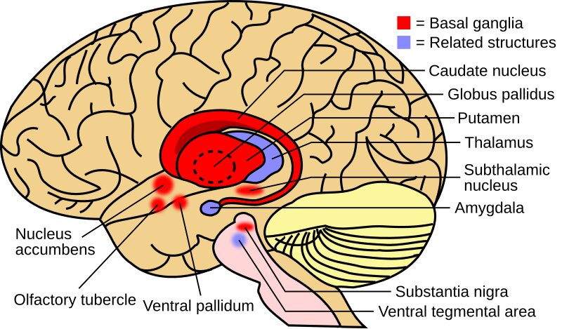
The basal ganglia are a group of nuclei located deep within the cerebral hemispheres, weighing about 40 grams and forming a major component of the motor control system. These structures include the caudate nucleus, putamen, and globus pallidus. Together, they help regulate voluntary motor movements, procedural learning, and routine behaviors like habits. The basal ganglia are also involved in cognitive and emotional functions, influencing how we initiate and inhibit actions. They work closely with the cerebral cortex and thalamus to modulate motor activity. Damage to this area can lead to movement disorders such as Parkinson’s disease or Huntington’s disease.
Amygdala
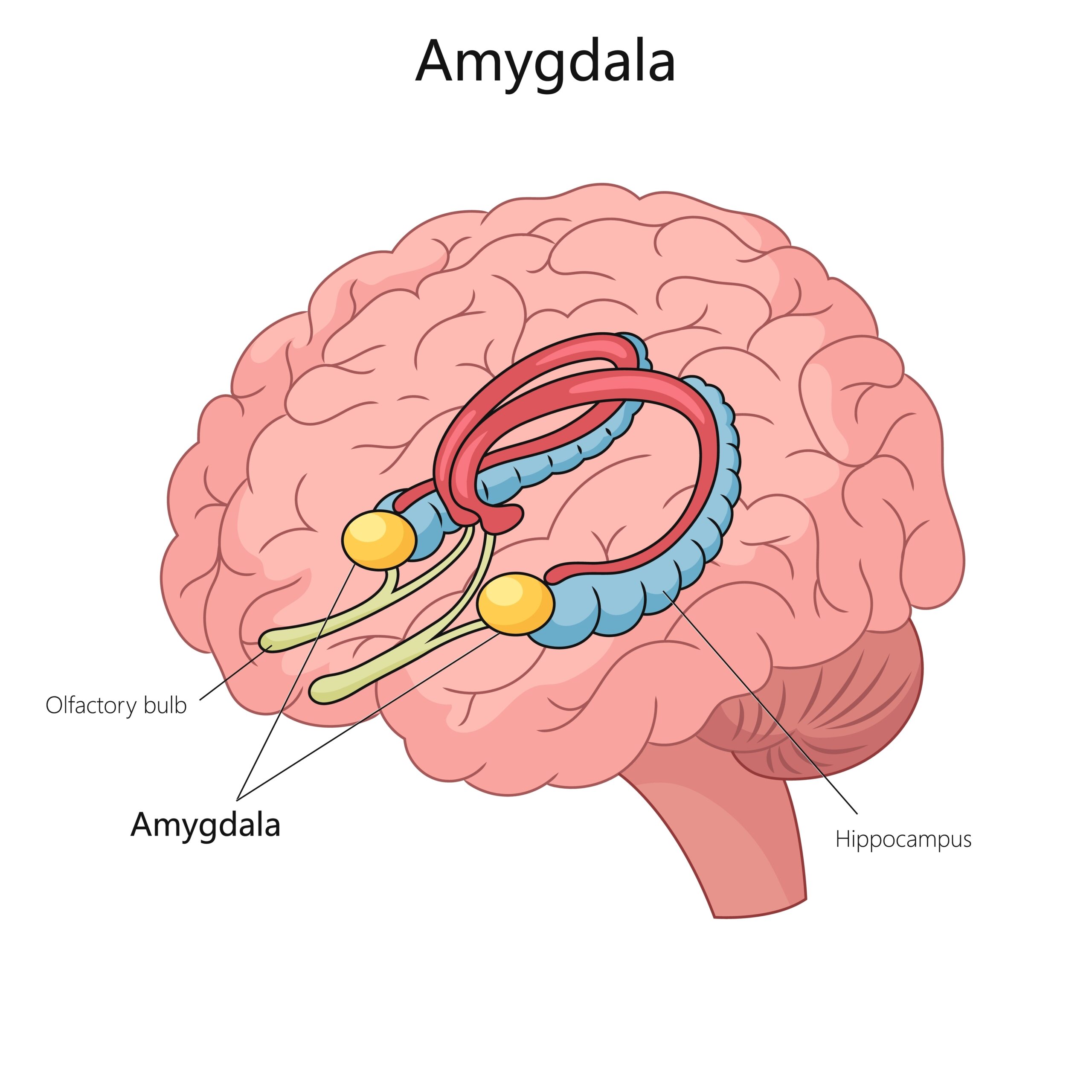
The amygdala is a small, almond-shaped structure located deep within the temporal lobes, approximately 1.5 centimeters in size. It is a key player in processing emotions, particularly fear and pleasure. The amygdala helps the brain identify potential threats and triggers the body’s fight-or-flight response. It is also involved in memory formation, especially emotional memories. The amygdala interacts with other parts of the brain, such as the hippocampus, to modulate stress responses. Its role in emotional regulation makes it essential for social behaviors. Dysfunction in the amygdala can lead to emotional dysregulation, anxiety, and mood disorders.
Hippocampus
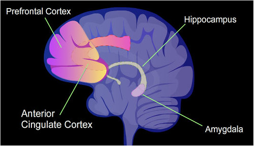
The hippocampus, a seahorse-shaped structure located in the temporal lobe, measures about 5 centimeters in length and is critical for memory formation. It is part of the limbic system and plays a significant role in converting short-term memories into long-term memories. The hippocampus also helps with spatial navigation and learning. Damage to this area can result in memory loss or difficulty forming new memories, as seen in conditions like Alzheimer’s disease. This structure interacts closely with the amygdala and prefrontal cortex in emotional memory processing. Its size and health are directly correlated with cognitive abilities, especially memory recall.
Pons
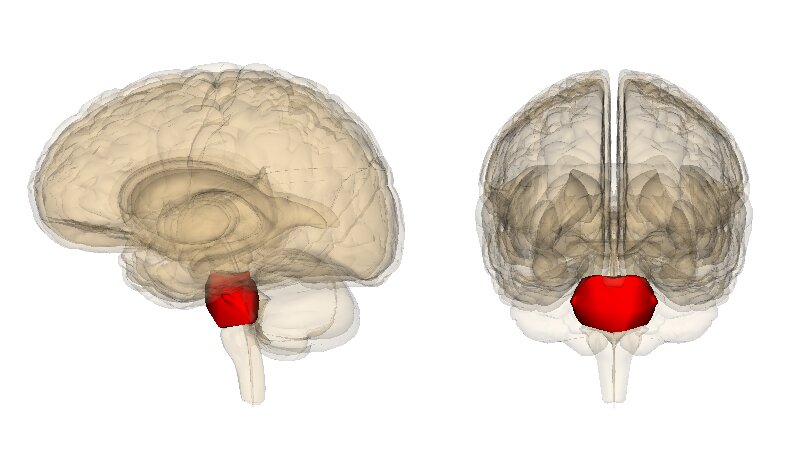
The pons is a bulbous structure located on the brainstem, about 2.5 centimeters in length and 25 grams in weight. It acts as a bridge between the cerebrum, cerebellum, and medulla oblongata. The pons contains nuclei that control vital functions such as breathing, sleep, and arousal. It also relays signals from the forebrain to the cerebellum, assisting in motor control and sensory analysis. Additionally, the pons is involved in regulating deep sleep and REM sleep cycles. Damage to this area can affect breathing, sleep patterns, and motor control. Its central location in the brainstem makes it essential for integrating many of the brain’s vital functions.
Medulla Oblongata
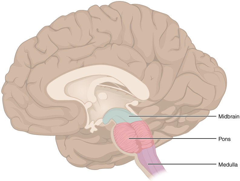
The medulla oblongata is located at the base of the brainstem, measuring about 3 centimeters in length, and it controls many of the body’s autonomic functions. It regulates vital involuntary activities such as heart rate, blood pressure, and breathing. The medulla also coordinates reflexes like coughing, sneezing, and swallowing. It connects the brain to the spinal cord, allowing for communication between the central nervous system and the body. Damage to the medulla can be life-threatening, as it can disrupt essential bodily functions. It works in conjunction with other brainstem structures to maintain homeostasis.
Corpus Callosum
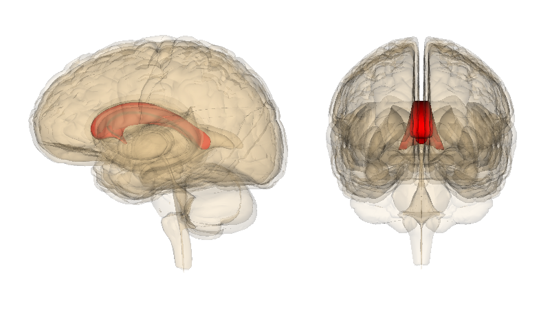
The corpus callosum is the largest white matter structure in the brain, spanning approximately 10 centimeters in length and weighing about 30 grams. It connects the left and right hemispheres, allowing for communication between both sides of the brain. This structure is essential for coordinating cognitive, motor, and sensory functions across hemispheres. The corpus callosum is made up of more than 200 million nerve fibers, facilitating the exchange of information between hemispheres. It also plays a role in integrating motor, sensory, and cognitive performances. Damage to the corpus callosum can result in split-brain syndrome, where the two hemispheres function independently. Its role in ensuring the hemispheres work cohesively is vital for overall brain function.
Ventricles
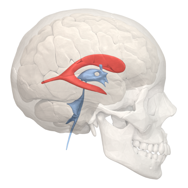
The brain’s ventricular system consists of four interconnected cavities filled with cerebrospinal fluid (CSF), which cushions and protects the brain. The largest are the lateral ventricles, followed by the third and fourth ventricles. These spaces are not solid structures but rather fluid-filled cavities that help remove waste products from the brain and transport nutrients. CSF flows through these ventricles, helping to maintain a stable environment for the brain’s delicate tissues. The ventricles play an essential role in protecting the brain from trauma by acting as a cushion against physical impacts. Enlarged ventricles can be a sign of neurological disorders such as hydrocephalus.
Pituitary Gland
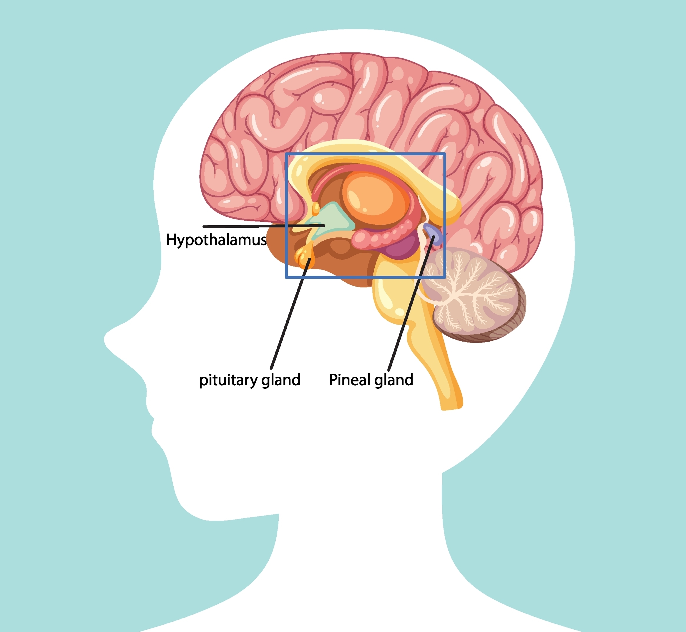
The pituitary gland, often referred to as the “master gland,” is about the size of a pea and is located at the base of the brain, beneath the hypothalamus. It weighs roughly 0.5 grams but plays a crucial role in regulating various hormones throughout the body. The pituitary gland is divided into anterior and posterior lobes, each responsible for secreting different hormones that control growth, metabolism, and reproduction. It receives signals from the hypothalamus to release or inhibit hormone production. Despite its small size, the pituitary gland influences almost every organ in the body. Damage to the pituitary gland can lead to hormonal imbalances and affect growth, thyroid function, and reproductive health.
Midbrain
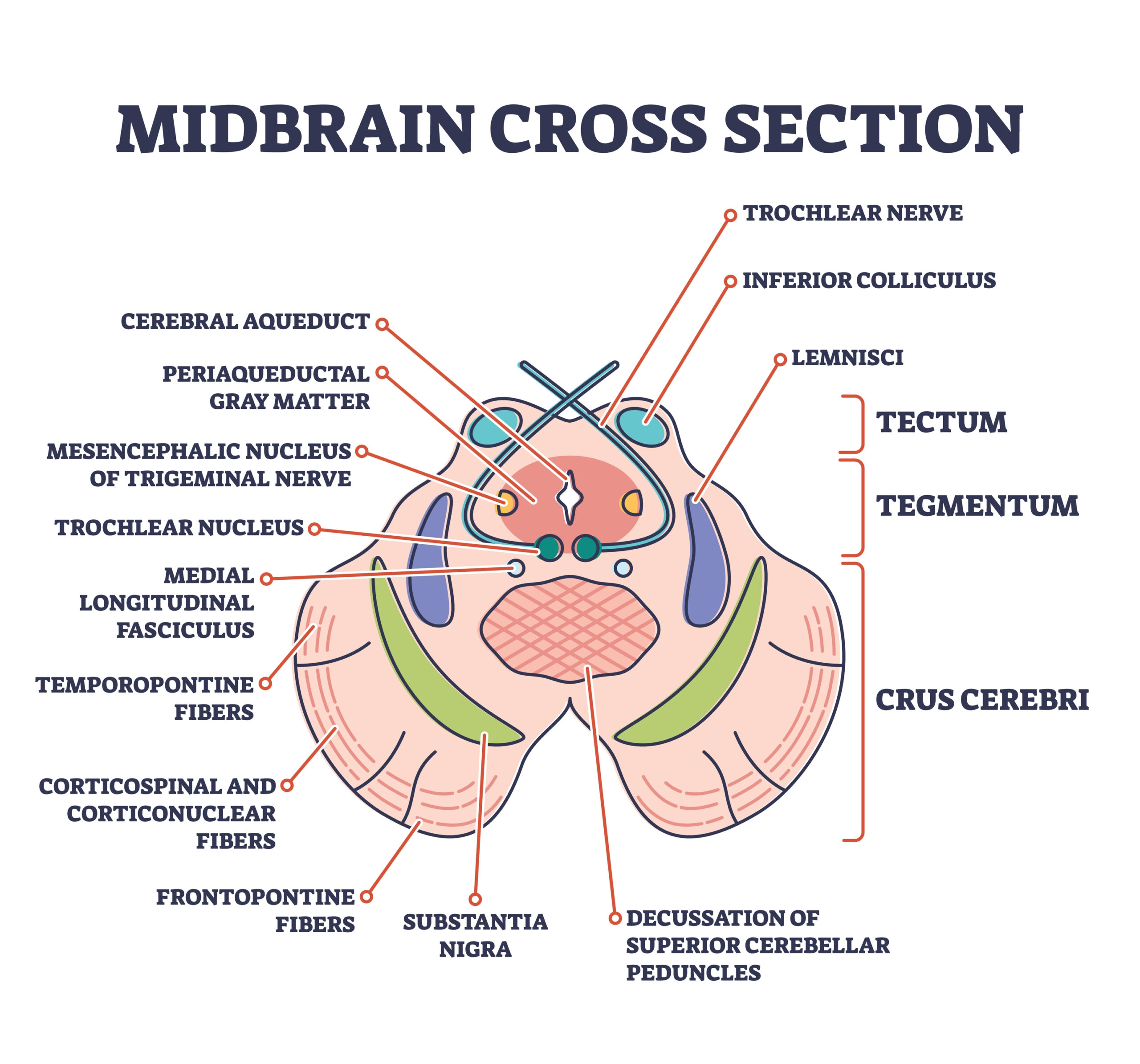
The midbrain, or mesencephalon, is located between the pons and the diencephalon, measuring about 2 centimeters in length. It is part of the brainstem and plays a role in motor movement, particularly eye movements, and auditory and visual processing. The midbrain contains structures like the superior and inferior colliculi, which are involved in visual and auditory reflexes, respectively. It also houses the substantia nigra, which is crucial for producing dopamine and controlling voluntary movements. The midbrain’s small size belies its importance in regulating these critical functions. Dysfunction in the midbrain, particularly in the substantia nigra, is linked to Parkinson’s disease.
Broca’s Area
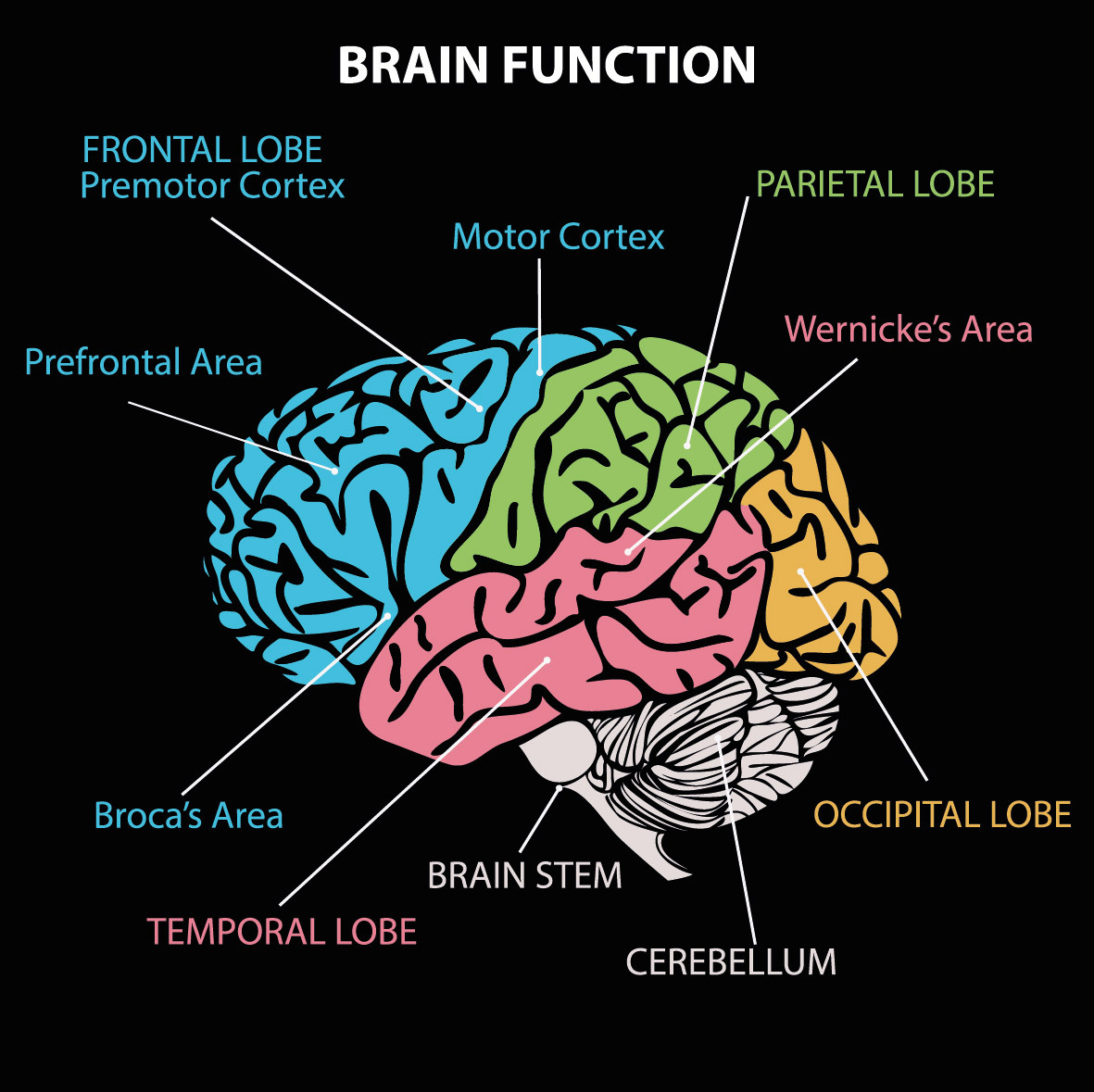
Broca’s area is a region in the frontal lobe, typically in the left hemisphere, associated with speech production. It is about 4 centimeters in size and plays a crucial role in language processing, including the articulation of words and sentences. Damage to Broca’s area can result in Broca’s aphasia, characterized by slow, halting speech and difficulty in forming complete sentences. This region coordinates with other parts of the brain to produce fluent speech and understand grammar. It works closely with Wernicke’s area, which is involved in language comprehension. Despite its relatively small size, Broca’s area is essential for communication.
Wernicke’s Area
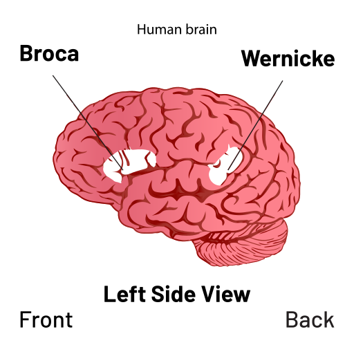
Wernicke’s area is located in the temporal lobe, typically in the left hemisphere, and measures about 4-5 centimeters in size. It is primarily responsible for language comprehension, enabling us to understand spoken and written language. Damage to Wernicke’s area can result in Wernicke’s aphasia, where individuals speak fluently but produce nonsensical or incoherent language. This region interacts with Broca’s area to ensure smooth language production and comprehension. Wernicke’s area is also involved in the processing of sounds related to speech. Its critical role in language makes it one of the most important areas for communication.
Pineal Gland
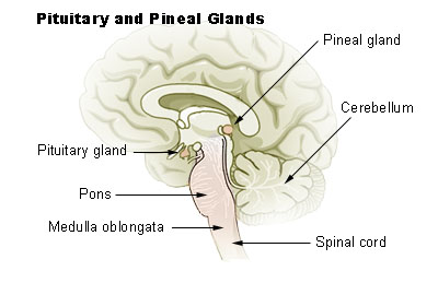
The pineal gland is a small, pinecone-shaped structure located near the center of the brain, measuring about 0.8 centimeters in length. It is part of the endocrine system and is responsible for producing melatonin, a hormone that regulates sleep-wake cycles. The pineal gland is sensitive to light, receiving signals from the retina to adjust melatonin production based on the time of day. This gland plays a vital role in maintaining circadian rhythms, which govern sleep patterns. Despite its small size, it has a significant impact on overall health and sleep quality. Dysfunction in the pineal gland can lead to sleep disorders and disruptions in circadian rhythms.
This article originally appeared on Rarest.org.
More From Rarest.Org
Migration is one of the most remarkable survival strategies in the animal kingdom. Every year, countless species travel vast distances, navigating through oceans, skies, and land to find food, breed, or escape harsh weather. Read more.
Retro advertising memorabilia is not only a window into the past but also a treasure trove for collectors. From iconic brand signs to old promotional items, these pieces have gained impressive value over time. Read more.
Majestic trees, some of the most iconic species on the planet, are now facing the threat of extinction. Human activities, environmental changes, and invasive species are pushing them to the brink. Read more.

